You have no items in your shopping cart.
Cart summary
Item 1 of 6
Item 1 of 6
AKT pS473 antibody
Catalog Number: orb420307

| Catalog Number | orb420307 |
|---|---|
| Category | Antibodies |
| Description | AKT pS473 antibody |
| Species/Host | Rabbit |
| Clonality | Polyclonal |
| Tested applications | DOT, ELISA, FC, IF, IHC, Multiplex Assay, WB |
| Reactivity | Human, Mouse, Rat |
| Isotype | IgG |
| Immunogen | Rabbit Anti-AKTpS473 Antibody was prepared by repeated immunizations in rabbits with a synthetic peptide corresponding to a C-terminus region near phospho Serine 473 of the human, mouse, rat and chicken AKT proteins conjugated to KLH. |
| Concentration | 1.0 mg/mL |
| Dilution range | ELISA: 1:15,000 - 1:60,000, FC: User Optimized, IHC: 1:100 - 1:500, IF: User Optimized, WB: 1:200 - 1:1000 |
| Form/Appearance | Liquid (sterile filtered) |
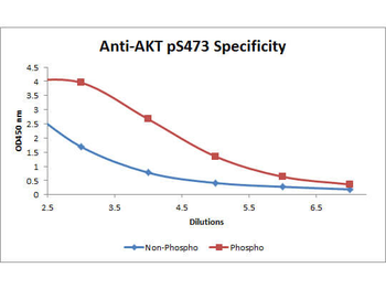
| Purity | This product was prepared from monospecific antiserum by immunoaffinity chromatography using a phospho peptide coupled to agarose beads followed by solid phase adsorption(s) against a non-phospho peptide and non-specific peptide to remove any unwanted reactivities. Assay by immunoelectrophoresis resulted in a single precipitin arc against anti-Rabbit Serum. This antibody is specific for phosphorylated human AKTpS473. Minimal reactivity occurs against non-phosphorylated AKT. Reactivity against AKT from other species may occur but has not yet been tested. |
| Conjugation | Unconjugated |
| UniProt ID | P31749 |
| NCBI | 62241011 |
| Storage | Store vial at -20° C or below prior to opening. This vial contains a relatively low volume of reagent (25 µL). To minimize loss of volume dilute 1:10 by adding 225 µL of the buffer stated above directly to the vial. Recap, mix thoroughly and briefly centrifuge to collect the volume at the bottom of the vial. Use this intermediate dilution when calculating final dilutions as recommended below. Store the vial at -20°C or below after dilution. Avoid cycles of freezing and thawing. |
| Buffer/Preservatives | 0.01% (w/v) Sodium Azide |
| Alternative names | rabbit anti-AKT pS473 Antibody, AKT1 phospho S473, Read more... |
| Note | For research use only |
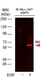
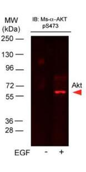
| Application notes | This AKT antibody is phospho specific for pS473 and is tested in ELISA, western blotting, immunohistochemistry (formalin-fixed paraffin-embedded sections), and immunofluorescence. By immunoblot a single band of the expected apparent molecular weight ~56kDa is observed. For immunohistochemistry no pre-treatment of sample is required. |
| Expiration Date | 12 months from date of receipt. |

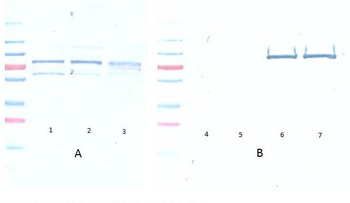
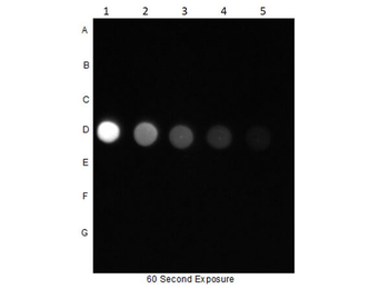
Dot Blot of Rabbit Anti-AKT pS473 Antibody. Dilutions in Columns: (1) 100 ng, (2) 33.33 ng, (3) 11.11 ng, (4) 3.7 ng, (5) 1.23 ng. Tested BSA Peptide Reactivity in Rows: (A) AKT1-BSA, (B) AKT1 pT308-BSA, (C) AKT1 S473-BSA, (D) AKT1 pS473-BSA, (E) CDC27 T244-BSA, (F) CDC27 pT244-BSA, (G) BSA control. Primary Antibody: Anti-AKTpS473 at 1 µg/mL overnight at 2-8°C. Secondary Antibody: Goat anti-Rabbit IgG HRP (p/n orb347654) at 1:70000 at RT for 30 mins. Block: BlockOut Buffer (p/n orb348644).
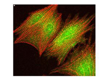
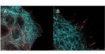
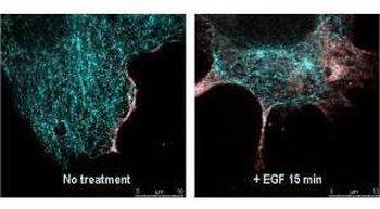
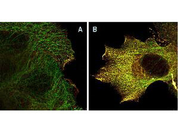
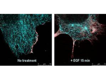
Immunofluorescence Confocal Microscopy of Rabbit anti-AKT pS473 antibody. Tissue: cardiomyocytes infected with adenovirus expressing with wild-type AKT. Fixation: 0.5% PFA. Antigen retrieval: not required. Primary antibody: AKT pS473 antibody at 1:40 for 1 h at RT. Secondary antibody: texas-red conjugated rabbit secondary antibody at 1:10000 for 45 min at RT. Localization: AKT pS473 is nuclear. Staining: AKT pS473 as green fluorescent signal with texas-red conjugated phalloidin (red) to label filamentous actin.
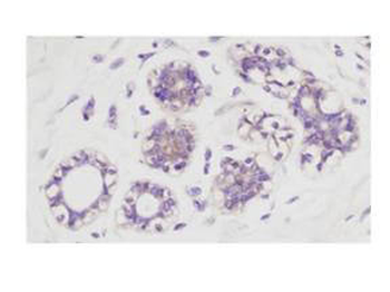
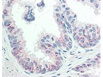
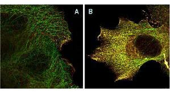
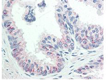
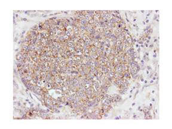
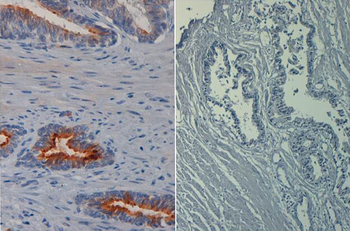
Immunohistochemistry at higher magnification of Rabbit Anti-Akt pS473 antibody. Tissue: human breast carcinoma. Fixation: formalin fixed paraffin embedded. Antigen retrieval: not required. Primary antibody: Akt pS473 antibody at 100 dilution for 1 h at RT. Secondary antibody: Dako's Techmate streptavidin-biotin reagents at 1:10000 for 45 min at RT. Localization: Akt pS473 is nuclear and occasionally cytoplasmic.
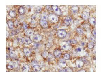
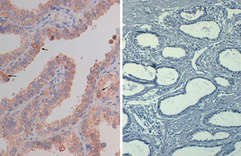
Immunohistochemistry of Rabbit anti-AKT pS473 antibody. Tissue: human breast carcinoma. Fixation: formalin fixed paraffin embedded. Antigen retrieval: not required. Primary antibody: AKT pS473 antibody at 1:100 for 1 h at RT. Secondary antibody: Dako's Techmate streptavidin-biotin reagents at 1:10000 for 45 min at RT. Localization: AKT pS473 is nuclear and occasionally cytoplasmic.

Immunohistochemistry of Rabbit Anti-Akt pS473 antibody. Tissue: human breast carcinoma. Fixation: formalin fixed paraffin embedded. Antigen retrieval: not required. Primary antibody: Akt pS473 antibody at 100 dilution for 1 h at RT. Secondary antibody: Dako's Techmate streptavidin-biotin reagents at 1:10000 for 45 min at RT. Localization: Akt pS473 is nuclear and occasionally cytoplasmic.

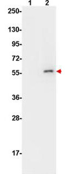
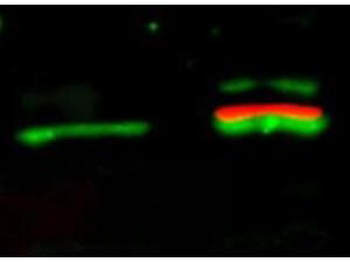
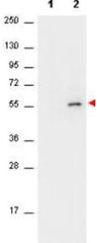
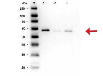
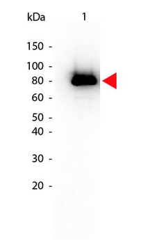
Western Blot of Rabbit anti-AKT pS473 antibody. Lane 1: AKT1 Recombinant Protein (p/n orb346473). Lane 2: AKT1 Mutant Human Recombinant Protein (p/n orb346474). Lane 3: AKT1 (phosphatase treated) Human Recombinant Protein (p/n orb346472). Load: 50 ng per lane. Primary antibody: AKT pS473 antibody at 1:1000 for overnight at 4°C. Secondary antibody: Peroxidase rabbit secondary antibody (p/n orb347654) at 1:40000 for 30 min at RT. Block: Blocking Buffer for Fluorescent Western Blotting (orb348637) for 30 min at RT. Predicted/Observed size: ~56 kDa for AKTpS473.
- Item 1 of 9
Akt (phospho-S473) antibody [orb344403]
ELISA, FC, IF, IHC, IP, Multiplex Assay, WB
Human, Monkey, Mouse, Rat
Mouse
Monoclonal
Unconjugated
100 μg - Item 1 of 9
Akt (phospho-S473) antibody [orb344404]
ELISA, FC, IF, IHC, IP, Multiplex Assay, WB
Human, Monkey, Mouse, Rat
Mouse
Monoclonal
Unconjugated
25 μl - Item 1 of 7
Akt (phospho-S473) antibody [orb344456]
ELISA, FC, IF, IHC, WB
Human, Mouse
Mouse
Monoclonal
Unconjugated
1 mg - Item 1 of 6
AKT pS473 antibody [orb345378]
DOT, ELISA, IF, IHC, Multiplex Assay, WB
Human, Mouse, Rat
Rabbit
Polyclonal
Unconjugated
100 μg - Item 1 of 4
AKT pS473 antibody (Biotin) [orb344541]
ELISA, FC, IF, IHC, IP, WB
Human, Monkey, Mouse, Rat
Mouse
Monoclonal
Biotin
50 μg


Submit a review
Filter by Rating

- 5 stars

- 4 stars
- 3 stars
- 2 stars
- 1 stars
