You have no items in your shopping cart.
Cart summary
Item 1 of 8
Item 1 of 8
AKT (phospho-T308) antibody
Catalog Number: orb420308

| Catalog Number | orb420308 |
|---|---|
| Category | Antibodies |
| Description | AKT (phospho-T308) antibody |
| Species/Host | Rabbit |
| Clonality | Polyclonal |
| Tested applications | DOT, ELISA, FC, IHC, WB |
| Reactivity | Human, Mouse, Rat |
| Isotype | IgG |
| Immunogen | Anti-AKT pT308 polyclonal antibody was produced by repeated immunizations with a phosphorylated synthetic peptide corresponding to residues surrounding threonine 308 of human AKT1 protein. |
| Concentration | 1.0 mg/mL |
| Dilution range | ELISA: 1:15,000, FC: User Optimized, IHC: 1:200, WB: User Optimized |
| Form/Appearance | Liquid (sterile filtered) |
| Purity | AKT phospho T308 Antibody was prepared from monospecific antiserum by immunoaffinity chromatography using phospho peptide coupled to agarose beads followed by solid phase adsorption(s) against non-phospho peptide and non-specific peptide to remove any unwanted reactivities. Assay by immunoelectrophoresis resulted in a single precipitin arc against anti-Rabbit Serum. This antibody is specific for phosphorylated human AKT. Minimal reactivity occurs against non-phosphorylated AKT. Reactivity against AKT from other species may occur but has not yet been tested. |
| Conjugation | Unconjugated |
| UniProt ID | P31749 |
| NCBI | 62241011 |
| Storage | Store vial at -20° C or below prior to opening. This vial contains a relatively low volume of reagent (25 µL). To minimize loss of volume dilute 1:10 by adding 225 µL of the buffer stated above directly to the vial. Recap, mix thoroughly and briefly centrifuge to collect the volume at the bottom of the vial. Use this intermediate dilution when calculating final dilutions as recommended below. Store the vial at -20°C or below after dilution. Avoid cycles of freezing and thawing. |
| Buffer/Preservatives | 0.01% (w/v) Sodium Azide |
| Alternative names | rabbit anti-AKT pT308 Antibody, AKT1 phospho T308, Read more... |
| Note | For research use only |
| Application notes | AKT antibody is phospho specific for pT308 and is tested for western blotting, immunohistochemistry, dot blot, and ELISA. This antibody is suitable for Flow Cytometry. |
| Expiration Date | 12 months from date of receipt. |
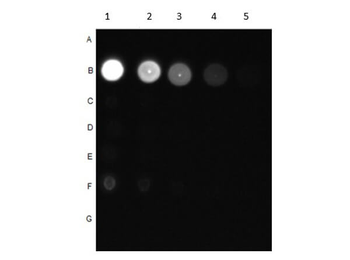
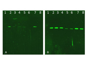
Dot Blot of Rabbit Anti-AKT pT308 Antibody. Dilutions in Columns: (1) 100 ng, (2) 33.33 ng, (3) 11.11 ng, (4) 3.7 ng, (5) 1.23 ng. Tested BSA Peptide Reactivity in Rows: (A) AKT1-BSA, (B) AKT1 pT308-BSA, (C) AKT1 S473-BSA, (D) AKT1 pS473-BSA, (E) CDC27 T244-BSA, (F) CDC27 pT244-BSA, (G) BSA control. Primary Antibody: Anti-AKT pT308 at 1 µg/mL overnight at 2-8°C. Secondary Antibody: Goat anti-Rabbit IgG HRP (p/n orb347654) at 1:70000 at RT for 30 mins. Block: BlockOut Buffer (p/n orb348644).

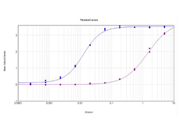
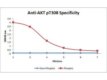
ELISA Results of Rabbit Anti-AKT pT308 Antibody tested against BSA-conjugated non-phospho [purple] and phospho [blue] forms of immunizing peptide. Each well was coated in duplicate with either 0.1 µg of conjugate. The working dilution is 1:81300. The starting dilution of antibody was 5 µg/ml and the X-axis represents the Log10 of a 3-fold dilution. This titration is a 4-parameter curve fit where the IC50 is defined as the titer of the antibody. Assay performed using HRP conjugate stabilizer, Goat Anti-Rabbit HRP conjugated (p/n orb347654) and TMB substrate (p/n orb348651).
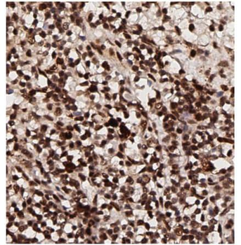
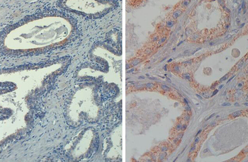
Immunohistochemistry of Rabbit Anti-AKT pT308 Antibody. Tissue: human breast tissue (lymph nodes). Antigen retrieval: HIER using citrate buffer for 20 minutes. Fixative: None. Primary Antibody: Anti-AKT phosphoT308 at 1:200 for 30 minutes at RT. Secondary Antibody: Anti-Rabbit Poly-HRP-IgG Ready to Use for 8 minutes at RT. Counterstain: Hematoxylin. Substrate: DAB. Analysis: Strong staining in nucleus. May be suitable with more dilutions.

Immunohistochemistry of Rabbit Anti-AKT pT308 Antibody. Tissue: human lung tissue. Antigen retrieval: HIER using citrate buffer for 20 minutes. Fixative: None. Primary Antibody: Anti-AKT phosphoT308 at 1:200 for 30 minutes at RT. Secondary Antibody: Anti-Rabbit Poly-HRP-IgG Ready to Use for 8 minutes at RT. Counterstain: Hematoxylin. Substrate: DAB. Analysis: Strong staining in nucleus. May be suitable with more dilutions.
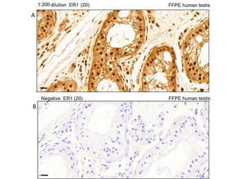
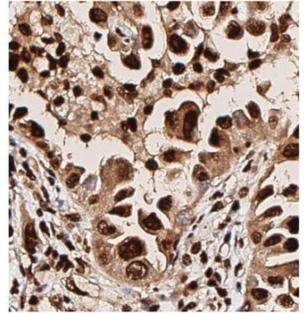
Immunohistochemistry of Rabbit Anti-AKT pT308 Antibody. Tissue: human testis tissue. Antigen retrieval: Heat induced antigen retrieval was performed using Leica Bond Epitope Retrieval Buffer 1 (Citrate solution, pH6.0) for 20 minutes. Fixative: None. Primary Antibody: (A). Anti-AKTpT308 at 1:200 for 30 minutes at RT. (B). Negative control. Secondary Antibody: Anti-Rabbit Poly-HRP-IgG Ready to Use for 8 minutes at RT. Counterstain: Hematoxylin. Substrate: DAB. Analysis: Cells in seminiferous ducts and Leydig cells show moderate cytoplasmic staining.
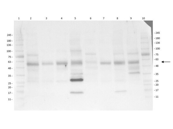
Multi-Lysate Western Blot of Rabbit Anti-AKT pT308 Antibody. Lane 1: Opal Pre-stained Molecular Weight Marker. Lane 2: Human Spleen Lysate. Lane 3: Hu Small Intestine Lysate. Lane 4: Hu Placenta Lysate. Lane 5: Hu Skeletal Muscle Lysate. Lane 6: Hu Brain Cerebellum Lysate. Lane 7: Hu Lung Lysate. Lane 8: Hu Tonsil Lysate. Lane 9: Hu Thymus Lysate. Lane 10: Opal Pre-stained Molecular Weight Marker. Primary Antibody: Anti-AKT pT308 at 1:1000 overnight at 2-8°C. Secondary Antibody: Goat Anti-Rabbit IgG HRP (p/n orb347654) at 1:40000 at RT for 60mins. Block: BlockOut Buffer (p/n orb348644). Predicted MW: ~55 kDa. Observed MW: ~28, ~58 kDa. Notes: Ubiquitous.
Western Blot of Rabbit AKT Antibodies. Lane 1: NIR MW protein ladder. Lane 2: AKT1, recombinant: orb346473. Lane 3: AKT1, phosphatase-treated: orb346472. Lane 4: AKT1, mutant T308A/S473A: orb346474. Lane 5: AKT2, recombinant: orb346475. Lane 6: AKT2, phosphatase-treated: orb346470. Lane 7: AKT3, recombinant: orb346476. Lane 8: AKT3, phosphatase-treated: orb346471. Load: 50 ng per lane. Blot A: orb345379 Anti-Akt pT308 used at 1:2270, Blot B: orb750474 Anti-Akt used 1:1000.

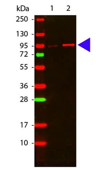
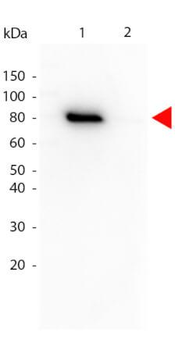
Western Blot of Rabbit anti-Akt phospho T308 antibody. Lane 1: GST tagged AKT1 un-active recombinant protein. Lane 2: GST tagged AKT1 active recombinant protein. Load: 50 ng per lane. Primary antibody: Akt phospho T308 antibody at 1:1000 for overnight at 4°C. Secondary antibody: DyLight™ 649 rabbit secondary antibody at 1:20000 for 30 min at RT. Block: orb348637 for 30 min at RT. Other band(s): none.
- Item 1 of 8
Akt (phospho-T308) antibody [orb345379]
DOT, ELISA, FC, IHC, WB
Human, Mouse, Rat
Rabbit
Polyclonal
Unconjugated
100 μg - Item 1 of 5
AKT1 (phospho-Thr308/Thr309) antibody [orb6780]
FC, ICC, IHC-P, WB
Bovine, Canine, Gallus, Porcine, Rabbit, Sheep
Human, Mouse, Rat
Rabbit
Polyclonal
Unconjugated
100 μl, 50 μl, 200 μl - Item 1 of 3
AKT (phospho-T308) antibody [orb213539]
IH, WB
Human, Mouse, Rat, Zebrafish
Rabbit
Polyclonal
Unconjugated
200 μl, 100 μl, 30 μl - Item 1 of 3
AKT pT308 antibody (Biotin) [orb344542]
DOT, ELISA, FC, IHC, IP, WB
Human, Monkey, Mouse, Rat
Mouse
Monoclonal
Biotin
50 μg - Item 1 of 3
AKT (phospho-Thr308) antibody [orb14786]
FC, IHC-P, WB
Bovine, Canine, Gallus, Porcine, Rabbit, Sheep
Human, Mouse, Rat
Rabbit
Polyclonal
Unconjugated
200 μl, 50 μl, 100 μl


Submit a review
Filter by Rating

- 5 stars

- 4 stars
- 3 stars
- 2 stars
- 1 stars
