You have no items in your shopping cart.
Cart summary
Item 1 of 6
Item 1 of 6
AIFM1 Antibody
Catalog Number: orb1248086

| Catalog Number | orb1248086 |
|---|---|
| Category | Antibodies |
| Description | AIFM1 Antibody |
| Species/Host | Goat |
| Clonality | Polyclonal |
| Tested applications | ELISA, IF, WB |
| Predicted Reactivity | Bovine, Canine |
| Reactivity | Human, Mouse, Porcine, Rat |
| Immunogen | The immunogen for this antibody is: C-NEVAKLFNIHED |
| Concentration | 500 ug/mL |
| Dilution range | Peptide ELISA: antibody detection limit dilution 1:128000.Western Blot:Approx 70kDa band observed in lysates of cell line Jurkat and NIH3T3, and in Mouse and Rat Heart and Kidney lysates. Approx. 65kDa observed in Pig Heart lysates (calculated MW of 66.9kDa according to Human NP_004199.1 and 66.8kDa according to Mouse NP_036149.1, Rat NP_112646.1 and Pig NP_001284561.1). Recommended concentration: 0.01-0.1ug/ml. Primary incubation 1 hour at room temperature.Immunofluorescence: Strong expression of the protein seen in the Mitochondria of HeLa and U2OS cells. Recommended concentration: 10ug/ml. |
| Form/Appearance | Liquid |
| Conjugation | Unconjugated |
| MW | Approx 70kDa |
| Target | AIFM1 |
| UniProt ID | O95831 |
| NCBI | NP_001124318.1, NP_665811.1, NP_004199.1, NP_665812.1 |
| Storage | Aliquot and store at -20°C. Minimize freezing and thawing. |
| Buffer/Preservatives | Supplied at 0.5 mg/ml in Tris saline, 0.02% sodium azide, pH 7.3 with 0.5% bovine serum albumin. Aliquot and store at -20°C. Minimize freezing and thawing. |
| Alternative names | AIFM1, apoptosis-inducing factor, mitochondrion-as Read more... |
| Note | For research use only |
| Application notes | Peptide ELISA: antibody detection limit dilution 1:128000.Western Blot:Approx 70kDa band observed in lysates of cell line Jurkat and NIH3T3, and in Mouse and Rat Heart and Kidney lysates. Approx. 65kDa observed in Pig Heart lysates (calculated MW of 66.9kDa according to Human NP_004199.1 and 66.8kDa according to Mouse NP_036149.1, Rat NP_112646.1 and Pig NP_001284561.1). Recommended concentration: 0.01-0.1ug/ml. Primary incubation 1 hour at room temperature.Immunofluorescence: Strong expression of the protein seen in the Mitochondria of HeLa and U2OS cells. Recommended concentration: 10ug/ml. |
| Expiration Date | 12 months from date of receipt. |
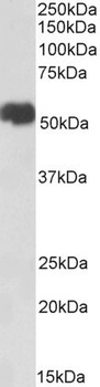
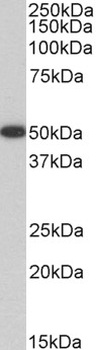
orb1248086 (0.01 ug/ml) staining of Jurkat lysate (35 ug protein in RIPA buffer). Detected by chemiluminescence.
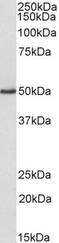
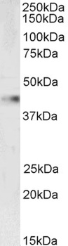
orb1248086 (0.1 ug/ml) staining of NIH3T3 lysate (35 ug protein in RIPA buffer) Detected by chemiluminescence.
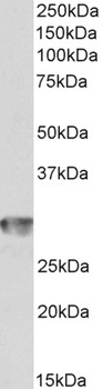
orb1248086 (0.01 ug/ml) staining of Mouse (A), Rat (B) and Pig (C) Heart lysate (35 ug protein in RIPA buffer). Detected by chemiluminescence.
orb1248086 (0.03 ug/ml) staining of Mouse (A) and Rat (B) Kidney lysate (35 ug protein in RIPA buffer). Detected by chemiluminescence
orb1248086 Immunofluorescence analysis of paraformaldehyde fixed HeLa cells, permeabilized with 0.15% Triton. Primary incubation 1hr (10 ug/ml) followed by Alexa Fluor 488 secondary antibody (4 ug/ml), showing Mitochondrial staining.
orb1248086 Immunofluorescence analysis of paraformaldehyde fixed U2OS cells, permeabilized with 0.15% Triton. Primary incubation 1hr (10 ug/ml) followed by Alexa Fluor 488 secondary antibody (4 ug/ml), showing Mitochondrial staining.
- Item 1 of 11
AIFM1 Antibody [orb1239169]
ELISA, IHC-P, WB
Human, Mouse, Rat
Rabbit
Polyclonal
Unconjugated
0.1 mg, 0.02 mg - Item 1 of 10
- Item 1 of 9
- Item 1 of 9
AIF/AIFM1 Antibody [orb251549]
FC, ICC, IF, IHC, WB
Hamster
Human, Mouse, Rat
Rabbit
Polyclonal
Unconjugated
10 μg, 100 μg - Item 1 of 8
AIFM1 Antibody [orb1239191]
ELISA, IF, WB
Mouse, Rat
Human
Rabbit
Polyclonal
Unconjugated
0.1 mg, 0.02 mg


Submit a review
Filter by Rating

- 5 stars

- 4 stars
- 3 stars
- 2 stars
- 1 stars
