You have no items in your shopping cart.
Cart summary
Item 1 of 8
Item 1 of 8
AHSP antibody
Catalog Number: orb750521

| Catalog Number | orb750521 |
|---|---|
| Category | Antibodies |
| Description | AHSP antibody |
| Species/Host | Rabbit |
| Clonality | Polyclonal |
| Tested applications | ELISA, IHC, WB |
| Reactivity | Human, Mouse |
| Isotype | Antiserum |
| Immunogen | Anti-AHSP Antibody was produced from whole rabbit serum prepared by repeated immunizations with the full length human AHSP protein. |
| Concentration | 115 mg/mL |
| Dilution range | ELISA: 1:2,000 - 1:10,000, IHC: 1:8000, WB: 1:500-1:1000 |
| Form/Appearance | Liquid (sterile filtered) |
| Purity | Anti-AHSP Antibody is directed against the human AHSP protein. The product was prepared from monospecific antiserum by delipidation and defibrination. A BLAST analysis was used to suggest cross reactivity with human and mouse. Cross-reactivity with AHSP from other sources have not been determined. |
| Conjugation | Unconjugated |
| UniProt ID | Q9NZD4 |
| NCBI | NP_057717.1 |
| Storage | Store vial at -20° C or below prior to opening. This vial contains a relatively low volume of reagent (25 µL). To minimize loss of volume dilute 1:10 by adding 225 µL of the buffer stated above directly to the vial. Recap, mix thoroughly and briefly centrifuge to collect the volume at the bottom of the vial. Use this intermediate dilution when calculating final dilutions as recommended below. Store the vial at -20°C or below after dilution. Avoid cycles of freezing and thawing. |
| Buffer/Preservatives | 0.01% (w/v) Sodium Azide |
| Alternative names | rabbit anti-AHSP antibody, Alpha-hemoglobin-stabil Read more... |
| Note | For research use only |
| Application notes | Anti-AHSP Antibody has been tested in western blot and immunohistochemistry and is suitable for immunofluorescence microscopy using paraformaldehyde-fixed primary cardiomyocyte cultures and ELISA. Specific conditions for reactivity should be optimized by the end user. |
| Expiration Date | 12 months from date of receipt. |
Immunohistochemistry of Rabbit anti-AHSP antibody. AHSP (A) stains residual EPs and not lymphoma cells in DLBCL, whereas CD71 (B) stains both lymphoma cells and EPs. C. Corresponding H&E. AHSP (D) does not metastatic carcinoma, whereas CD71 (E) does. F. Corresponding H&E. Fixation: acetic acid-zinc-formalin and formalin fixation, embedded in paraffin. Antigen retrieval: TRIS-EDTA pH9.0. Primary antibody: AHSP antibody at 1:8000 for overnight at 4°C Secondary antibody: anti-rabbit secondary at 1:10000 for 45 min at RT. Localization: Anti-AHSP is cytoplasmic. Staining: AHSP antibody as precipitated brown signal with a purple nuclear counterstain using Bond-max™ – fully automated for IHC.
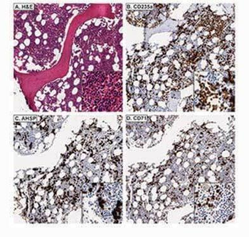
Immunohistochemistry of Rabbit anti-AHSP antibody. AHSP and CD71 stain megakaryocytes in primary myelofibrosis. Tissue: A.) Primary myelofibrosis, H&E. B.) CD235a stains both nucleated EPs and mature, anucleate RBCs. AHSP C.) and CD71. D.) variably stain megakaryocytes and also stain nucleated EPs. Fixation: acetic acid-zinc-formalin and formalin fixation, embedded in paraffin. Antigen retrieval: TRIS-EDTA pH9.0. Primary antibody: AHSP antibody at 1:8000 for overnight at 4°C Secondary antibody: anti-rabbit secondary at 1:10000 for 45 min at RT. Localization: Anti-AHSP is cytoplasmic. Staining: AHSP antibody as precipitated brown signal with a purple nuclear counterstain using Bond-max™ – fully automated for IHC.

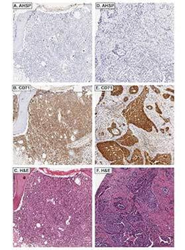
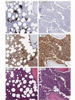
Immunohistochemistry of Rabbit anti-AHSP antibody. CD71 stains myeloid blasts in acute myeloid leukemia, whereas AHSP does not. AHSP stains residual EPs and not myeloid blasts in acute myeloid leukemia with minimal differentiation. (A), whereas CD71 stains both myeloid blasts and EPs (B). AHSP does not stain myeloid blasts in acute myelomonocytic leukemia (D), whereas CD71 does (E). C and F are corresponding H&Es, respectively. Fixation: acetic acid-zinc-formalin and formalin fixation, embedded in paraffin. Antigen retrieval: TRIS-EDTA pH9.0. Primary antibody: AHSP antibody at 1:8000 for overnight at 4°C Secondary antibody: anti-rabbit secondary at 1:10000 for 45 min at RT. Localization: Anti-AHSP is cytoplasmic. Staining: AHSP antibody as precipitated brown signal with a purple nuclear counterstain using Bond-max™ – fully automated for IHC.
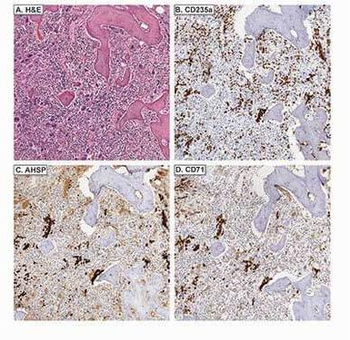
Immunohistochemistry of Rabbit anti-AHSP antibody. Tissue: A.) Acute erythroleukemia, H&E. B.) CD235a stains erythroid blasts and mature, anucleate RBCs. C.) AHSP stains erythroid blasts. D.) CD71 stains erythroid blasts. Fixation: acetic acid-zinc-formalin and formalin fixation, embedded in paraffin. Antigen retrieval: TRIS-EDTA pH9.0. Primary antibody: AHSP antibody at 1:8000 for overnight at 4°C. Secondary antibody: anti-rabbit secondary at 1:10000 for 45 min at RT. Localization: Anti-AHSP is cytoplasmic. Staining: AHSP antibody as precipitated brown signal with a purple nuclear counterstain using Bond-max™ – fully automated for IHC.
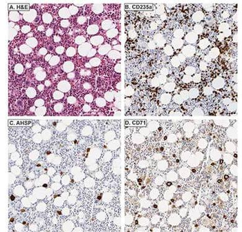
Immunohistochemistry of Rabbit anti-AHSP antibody. Tissue: A.) Giant pronormoblasts are evident in parvoviral infection, H&E. B.) CD235a does not stain giant pronormoblasts. C.) AHSP and D.) CD71 stain giant pronormoblasts. Fixation: acetic acid-zinc-formalin and formalin fixation, embedded in paraffin. Antigen retrieval: TRIS-EDTA pH9.0. Primary antibody: AHSP antibody at 1:8000 for overnight at 4°C Secondary antibody: anti-rabbit secondary at 1:10000 for 45 min at RT. Localization: Anti-AHSP is cytoplasmic. Staining: AHSP antibody as precipitated brown signal with a purple nuclear counterstain using Bond-max™ – fully automated for IHC.

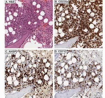
Immunohistochemistry of Rabbit anti-AHSP antibody. Tissue: A.) Normal bone marrow, H&E. B.) CD235a stains both nucleated EPs and mature, anucleate RBCs. C.) AHSP stains nucleated EPs, but not mature, anucleate RBCs. D.) CD71 stains nucleated EPs, but not mature, anucleate RBCs. Fixation: acetic acid-zinc-formalin and formalin fixation, embedded in paraffin. Antigen retrieval: TRIS-EDTA pH9.0. Primary antibody: AHSP antibody at 1:8000 for overnight at 4°C. Secondary antibody: anti-rabbit secondary at 1:10000 for 45 min at RT. Localization: Anti-AHSP is cytoplasmic. Staining: AHSP antibody as precipitated brown signal with a purple nuclear counterstain using Bond-max™ – fully automated for IHC.
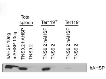
Western Blot of Rabbit anti-AHSP antibody. Lane 1: Recombinant hAHSP (10 ng). Lane 2: Recombinant mAHSP (10 ng). Lane 3: mice Spleen cells transfected with TNS9.2-hAHSP. Lane 4: mice Spleen cells transfected with TNS9.2 control vector. Lane 5: mice Spleen cells transfected with TNS9.2-hAHSP fractionated by MACS using Ter119+ microbeads. Lane 6: mice Spleen cells transfected with TNS9.2 control vector fractionated by Ter119+. Lane 7: mice Spleen cells transfected with TNS9.2-hAHSP fractionated by Ter119-. Lane 8: Spleen cells from mice transduced with TNS9.2 control vector fractionated by Ter119-. Load: 10 ng per lane. Primary antibody: AHSP antibody at 1:1000 for overnight at 4°C. Secondary antibody: HRP Streptavidin secondary antibody at 1:40000 for 30 min at RT. Block: 5% dry milk 30 min at RT. Predicted/Observed size: ~12 kDa. Other band(s): none.

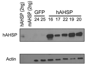
Western Blot of Rabbit anti-AHSP antibody. Lane 1: Recombinant hAHSP (2 ng). Lane 2: Recombinant mAHSP (2 ng). Lane 3: RBC Lysates Mouse #24 - GFP. Lane 4: RBC Lysates Mouse #25 - GFP. Lane 5: RBC Lysates Mouse #16 - hAHSP. Lane 6: RBC Lysates Mouse #17 - hAHSP. Lane 7: RBC Lysates Mouse #22 - hAHSP. Lane 8: RBC Lysates Mouse #19 - hAHSP. Lane 9: RBC Lysates Mouse #20 - hAHSP. Load: if not described differently, 10 ng per lane. Primary antibody: hAHSP antibody, Beta-Actin antibody at 1:1000 for overnight at 4°C. Secondary antibody: HRP Streptavidin secondary antibody at 1:40000 for 30 min at RT. Block: 5% dry milk 30 min at RT. Predicted/Observed size: ~12 kDa. Other band(s): none.
- Item 1 of 8
- Item 1 of 6
- Item 1 of 1
- Item 1 of 2
- Item 1 of 1


Submit a review
Filter by Rating

- 5 stars

- 4 stars
- 3 stars
- 2 stars
- 1 stars
