You have no items in your shopping cart.
Cart summary
Item 1 of 3
Item 1 of 3
AHA1 antibody
Catalog Number: orb345001

| Catalog Number | orb345001 |
|---|---|
| Category | Antibodies |
| Description | AHA1 antibody |
| Species/Host | Rat |
| Clonality | Monoclonal |
| Clone Number | 25F2.D9 |
| Tested applications | ELISA, IHC, WB |
| Reactivity | Human |
| Isotype | IgG2a |
| Immunogen | This Protein G purified monoclonal antibody was produced in rats by repeated immunizations with full length recombinant mouse AHA1 protein followed by hybridoma development. |
| Concentration | 1.0 mg/mL |
| Dilution range | ELISA: 1:20,000, IHC: 5-10 µg/mL, WB: 1:1,000 |
| Form/Appearance | Liquid (sterile filtered) |
| Purity | This product is an IgG fraction antibody purified from tissue culture supernatant by Protein-G chromatography, followed by extensive dialysis against the buffer stated above. It is directed against human Aha1 protein. A BLAST analysis was used to suggest cross-reactivity with Aha1 protein from mouse, human and chimpanzee based on 100% homology with the immunizing sequence. Reactivity against homologues from other sources is not known. |
| Conjugation | Unconjugated |
| UniProt ID | Q8BK64 |
| NCBI | NP_666148.1 |
| Storage | Store vial at -20° C prior to opening. Aliquot contents and freeze at -20° C or below for extended storage. Avoid cycles of freezing and thawing. Centrifuge product if not completely clear after standing at room temperature. This product is stable for several weeks at 4° C as an undiluted liquid. Dilute only prior to immediate use. |
| Buffer/Preservatives | None |
| Alternative names | rat anti-Aha1 Antibody, Aha1, Ahsa1 antibody, Acti Read more... |
| Note | For research use only |
| Application notes | This Protein-G purified antibody has been tested for use in ELISA, immunohistochemistry and western blotting. Specific conditions for reactivity should be optimized by the end user. Expect a band approximately 38-40 kDa in size corresponding to Aha1 protein by western blotting in the appropriate cell lysate or extract. |
| Expiration Date | 12 months from date of receipt. |
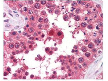
Biorbyt's anti-AHA1 monoclonal antibody was used at a 5-10 µg/mL to detect AHA1 in the seminiferous tubule of human testis (40X) showing moderate staining. Leydig cells showed faint to moderate staining. Expression of AHA1 is reported in many epithelial and lymphatic tissues, with cytoplasmic localization. This antibody showed moderate cytoplasmic staining of a variety of epithelial tissues and lymphoid organs such as spleen and tonsil with minimal background staining. The image shows the localization of the antibody as the precipitated red signal, with a hematoxylin purple nuclear counterstain. Tissue was formalin-fixed and paraffin embedded.
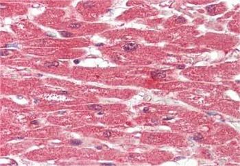
Immunohistochemistry of Rat anti-AHA1 antibody. Tissue: human heart. Fixation: formalin fixed paraffin embedded. Antigen retrieval: not required. Primary antibody: anti-AHA1 antibody at 10 µg/mL for 1 h at RT. Secondary antibody: Peroxidase rat secondary antibody at 1:10000 for 45 min at RT. Staining: AHA-1 as precipitated red signal with hematoxylin purple nuclear counterstain.
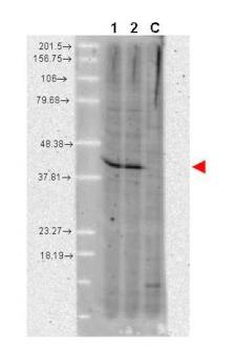
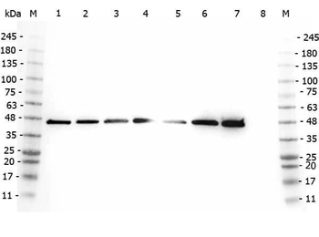
Western blot using Biorbyt's anti-AHA1 monoclonal antibody shows detection of a band ~42 kDa in size corresponding to AHA1. Molecular weight markers are shown at the left. Lane 1: A431 whole cell lysate (p/n orb348665) and Lane 2: MCF-7 whole cell lysate (p/n orb348664). A control lane is shown where primary antibody was omitted from the incubation (lane C). For best results, block the membrane overnight with 3% BSA in TBS followed by reaction with primary antibody diluted 1:1000 and use HRP conjugated anti-Rat IgG (p/n orb347790) secondary antibody diluted 1:20000 in blocking buffer (p/n orb348637) for detection.
- Item 1 of 10
AHA1/AHSA1 Antibody [orb1145840]
ELISA, FC, ICC, IF, IHC, WB
Human, Mouse, Rat
Rabbit
Polyclonal
Unconjugated
10 μg, 100 μg - Item 1 of 8
- Item 1 of 2
- Item 1 of 3
- Item 1 of 2


Submit a review
Filter by Rating

- 5 stars

- 4 stars
- 3 stars
- 2 stars
- 1 stars
