You have no items in your shopping cart.
Cart summary
Item 1 of 5
Item 1 of 5
ACVRL1 Antibody (N-term)
Catalog Number: orb1928871

| Catalog Number | orb1928871 |
|---|---|
| Category | Antibodies |
| Description | Purified Rabbit Polyclonal Antibody (Pab) |
| Species/Host | Rabbit |
| Clonality | Polyclonal |
| Clone Number | RB03736 |
| Tested applications | FC, IHC-P, WB |
| Reactivity | Human, Mouse |
| Isotype | Rabbit IgG |
| Antibody Type | Primary Antibody |
| Dilution range | WB: 1:1000, WB: 1:1000, WB: 1:1000, IHC-P: 1:50~100, FC: 1:10~50 |
| Form/Appearance | Purified polyclonal antibody supplied in PBS with 0.09% (W/V) sodium azide. This antibody is prepared by Saturated Ammonium Sulfate (SAS) precipitation followed by dialysis against PBS. |
| Conjugation | Unconjugated |
| MW | 56124 Da |
| Target | This ACVRL1 antibody is generated from rabbits immunized with a KLH conjugated synthetic peptide between 38-68 amino acids from the N-terminal region of human ACVRL1. |
| UniProt ID | P37023 |
| NCBI | NP_000011.2, NP_001070869.1 |
| Storage | Maintain refrigerated at 2-8°C for up to 2 weeks. For long term storage store at -20°C in small aliquots to prevent freeze-thaw cycles |
| Alternative names | Serine/threonine-protein kinase receptor R3, SKR3, Read more... |
| Note | For research use only |
| Expiration Date | 12 months from date of receipt. |

Western blot of ACVRL1 Pab. TOP LEFT: Mouse heart tissue lysate.
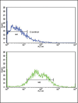
Flow cytometric analysis of HepG2 cells using ACVRL1 Antibody (N-term) (bottom histogram) compared to a negative control cell (top histogram). FITC-conjugated goat-anti-rabbit secondary antibodies were used for the analysis.
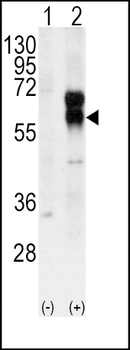
Western blot analysis of ACVRL1 (arrow) using rabbit polyclonal ACVRL1 Antibody (N-term). 293 cell lysates (2 ug/lane) either nontransfected (Lane 1) or transiently transfected with the ACVRL1 gene (Lane 2).
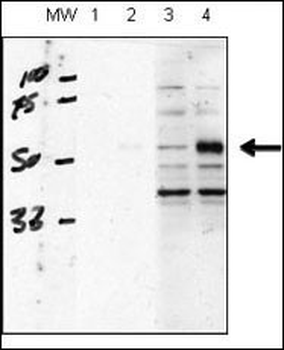
human chondrocytes (C28/I2 cells), transfected with empty vector (lane 1, 3) or ACVRL1 (lane 2, 4). RIPA lysis buffer, 20 ug/lane of protein, primary antibody dilution 1:1000, blocking solution is 5% milk in TBST (lane 1 and 2), 5% BSA in TBST (lane 3 and 4).
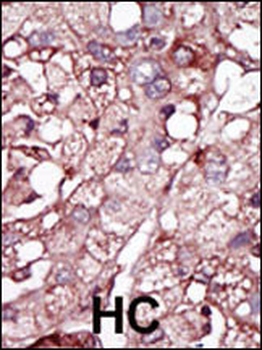
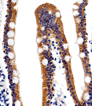
Formalin-fixed and paraffin-embedded human cancer tissue reacted with the primary antibody, which was peroxidase-conjugated to the secondary antibody, followed by DAB staining. This data demonstrates the use of this antibody for immunohistochemistry; clinical relevance has not been evaluated. BC = breast carcinoma; HC = hepatocarcinoma.
- Item 1 of 2

ACVRL1 Antibody (N-term) [orb1166291]
FC, IHC-P, WB
Human, Mouse
Rabbit
Polyclonal
Unconjugated
100 μl, 30 μl