You have no items in your shopping cart.
Cart summary
Item 1 of 16
Item 1 of 16

| Catalog Number | orb155559 |
|---|---|
| Category | Antibodies |
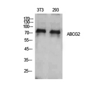
| Description | ABCG2 antibody |
| Species/Host | Rabbit |
| Clonality | Polyclonal |
| Tested applications | ICC, IF, IHC-P, WB |
| Isotype | IgG |
| Immunogen | KLH conjugated synthetic peptide derived from human ABCG2. Please contact us for the exact immunogen sequence. The peptide is available as orb12938. |
| Concentration | -100 μg (in 200 μl): 0.5 mg/ml-200 μg (in 400 μl): 0.5 mg/ml |
| Dilution range | IF/ICC: 1:100-500, IHC-P: 1:100-500, WB: 1:200-2000 |
| Form/Appearance | 10 mM PBS, 0.02% sodium azide |
| Purity | Polyclonal antibodies are purified by peptide affinity chromatography |
| Conjugation | Unconjugated |
| MW | 72 kDa |
| Target | ABCG2 |
| Entrez | 9429 |
| UniProt ID | Q9UNQ0, Q80W57, Q7TMS5 |
| Storage | Maintain refrigerated at 2-8°C for up to 2 weeks. For long term storage store at -20°C in small aliquots to prevent freeze-thaw cycles. |
| Alternative names | anti ABC15 antibody, anti ABCG2 antibody, anti ABC Read more... |
| Note | For research use only |
| Expiration Date | 12 months from date of receipt. |
Li, YiFu et al. MRP-1 and BCRP Promote the Externalization of Phosphatidylserine in Oxalate-treated Renal Epithelial Cells: Implications for Calcium Oxalate Urolithiasis Urology, 107, 271.e9-271.e17 (2017)
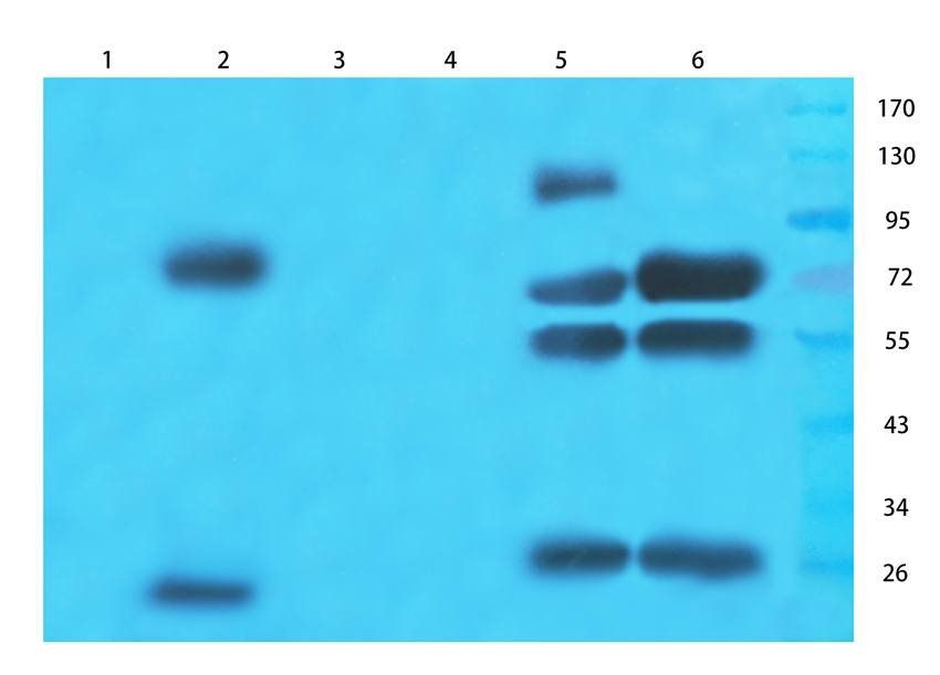
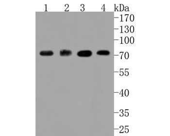
WB analysis of mouse brain (lane 1), rat small intestines (lane 2), cow milk (lane 3), hela cells (lane 4), human lung cancer (lane 5), human breast cancer (lane 6) using ABCG2 antibody (1 ug/ml)
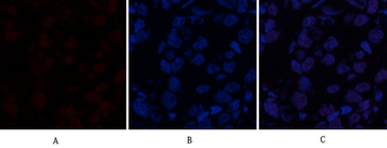
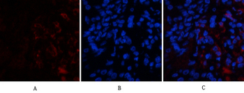
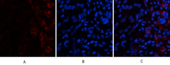
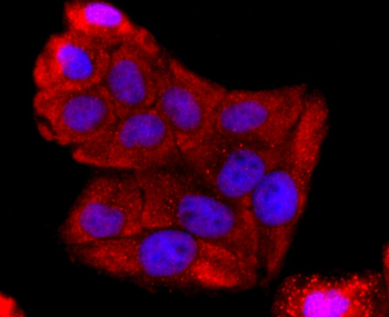
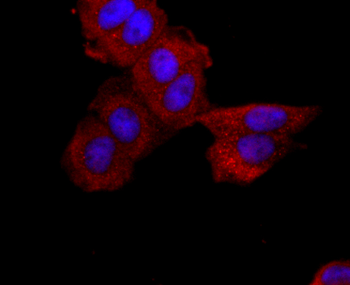
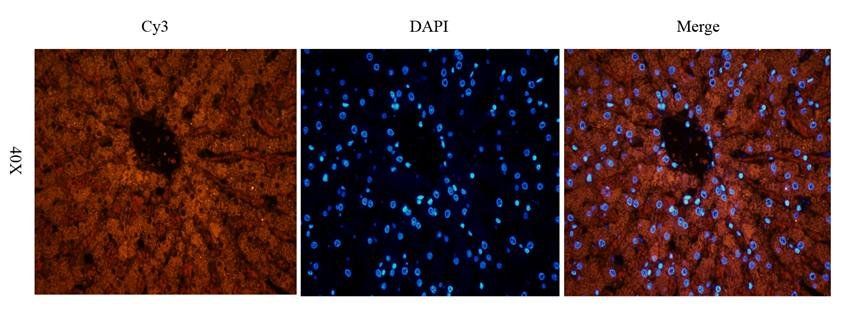
IF image of rat brain tissue using anti-ABCG2 (2.5 ug/ml)
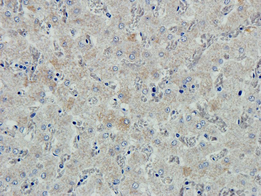
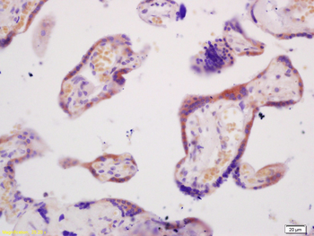
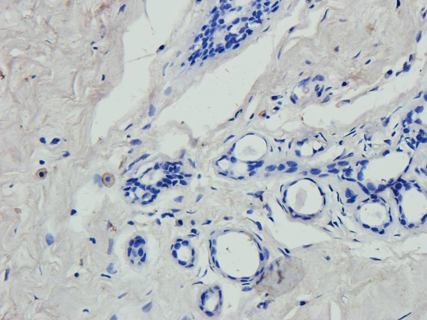
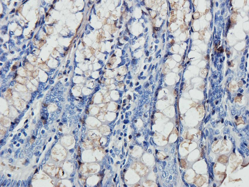
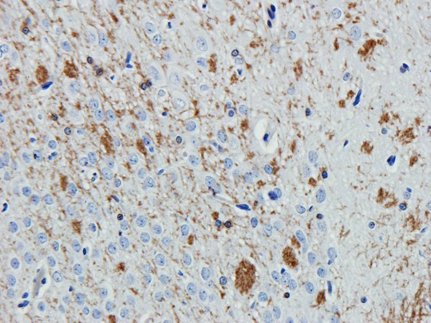
Immunohistochemical staining of paraffin embedded pig liver tissue using anti-ABCG2 (2.5 ug/ml)
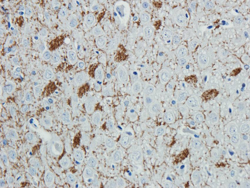
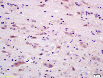
IHC-P image of rat brain tissue using ABCG2 antibody (2.5 ug/ml)
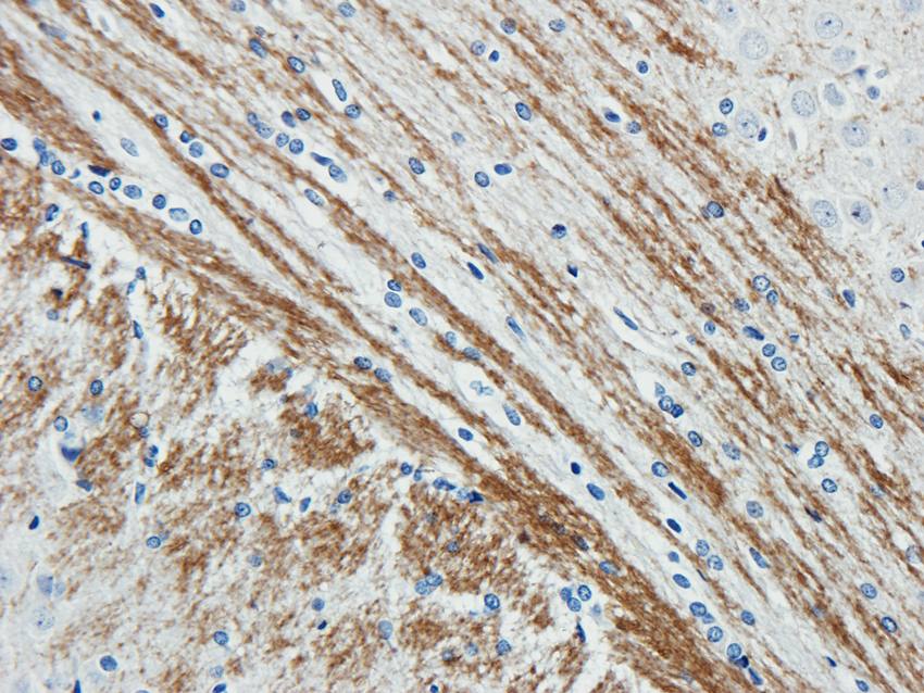
Immunohistochemical staining of rat brain tissue using ABCG2 antibody (2.5 ug/ml)
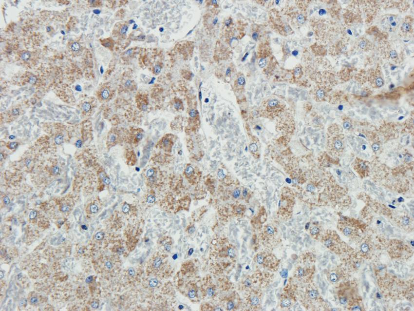
IHC-P staining of pig liver tissue using anti-ABCG2 (2.5 ug/ml)
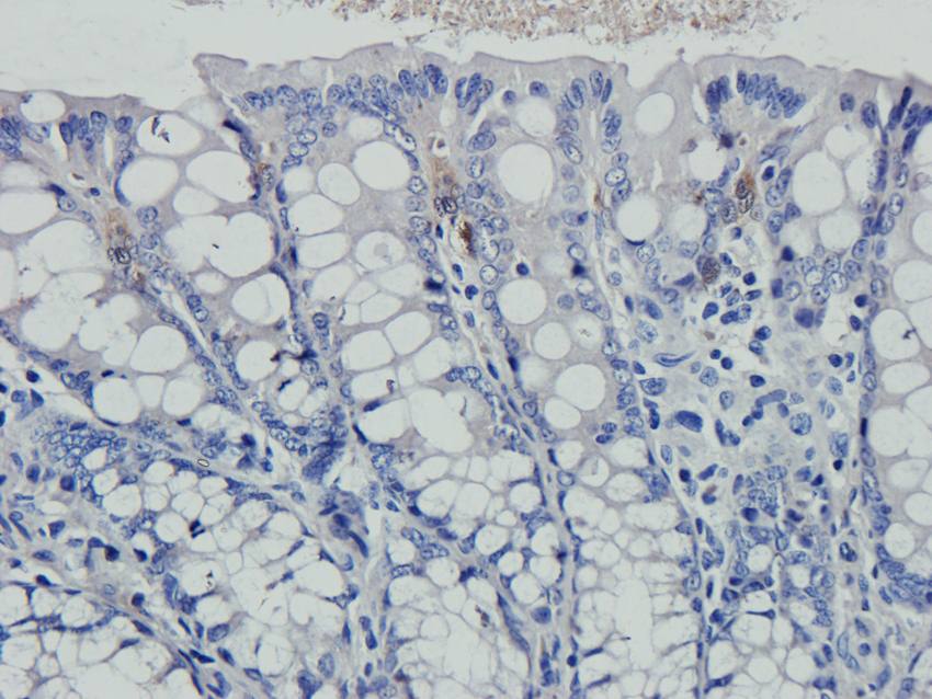
IHC-P image of guinea pig rectum tissue using anti-ABCG2 (2.5 ug/ml)
Immunohistochemical staining of paraffin embedded human breast tumour tissue using ABCG2 antibody (2.5 ug/ml)
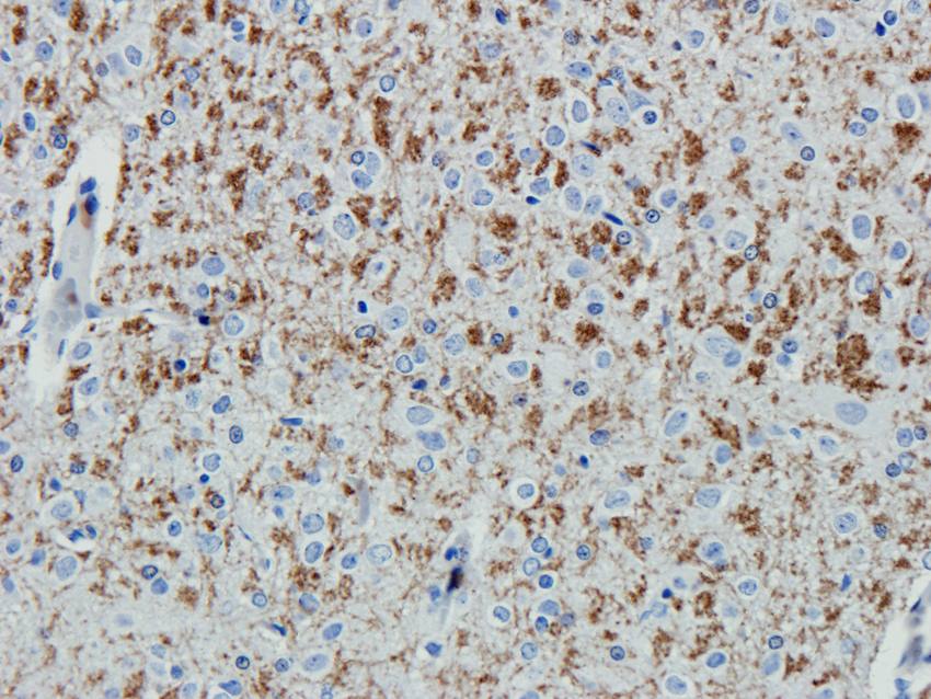
Immunohistochemical staining of rat brain tissue using ABCG2 antibody (2.5 ug/ml)
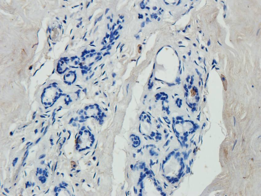
IHC-P staining of human breast tumour tissue using anti-ABCG2 (2.5 ug/ml)
IHC-P image of guinea pig rectum tissue using anti-ABCG2 (2.5 ug/ml)
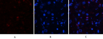
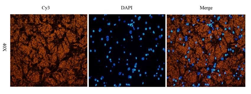
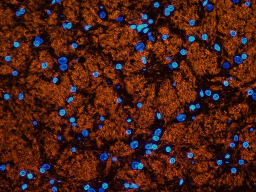
Immunofluorescence analysis of pig liver tissue using ABCG2 antibody (2.5 ug/ml)
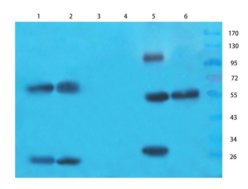
Western blot analysis of mouse brain (lane 1), rat small intestines (lane 2), cow milk (lane 3), hela cells (lane 4), human lung cancer (lane 5), human breast cancer (lane 6) using ABCG2 antibody (1 ug/ml)
Immunofluorescence image of rat brain tissue using ABCG2 antibody (2.5 ug/ml)
IHC-P staining of rat brain tissue using ABCG2 antibody (2.5 ug/ml)
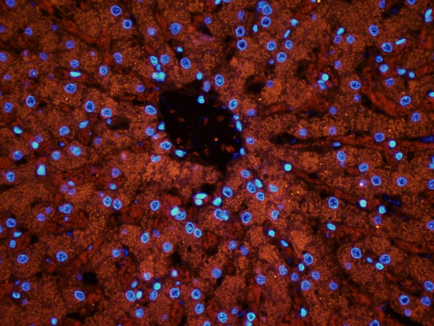
Immunofluorescence image of pig liver tissue using anti-ABCG2 (2.5 ug/ml)
- Item 1 of 7
- Item 1 of 3
ABCG2 Rabbit Polyclonal Antibody [orb312148]
ELISA, IF, IHC-Fr, IHC-P, WB
Mouse, Rat
Human, Mouse, Rat
Rabbit
Polyclonal
Unconjugated
50 μl, 100 μl - Item 1 of 5
- Item 1 of 4
ABCG2 Recombinant Rabbit Monoclonal Antibody [orb1499376]
IF, IHC-Fr, IHC-P, WB
Human, Mouse
Human
Rabbit
Recombinant
Unconjugated
50 μl, 100 μl - Item 1 of 4
ABCG2 rabbit pAb [orb767163]
ELISA, IF, IHC-P, WB
Human, Mouse, Rat
Polyclonal
Unconjugated
100 μl, 50 μl