You have no items in your shopping cart.
Cart summary
Item 1 of 4
Item 1 of 4
ABCB5 antibody
Catalog Number: orb345620

| Catalog Number | orb345620 |
|---|---|
| Category | Antibodies |
| Description | ABCB5 antibody |
| Species/Host | Rabbit |
| Clonality | Polyclonal |
| Tested applications | ELISA, IHC, WB |
| Reactivity | Human |
| Isotype | IgG |
| Immunogen | This affinity purified antibody was prepared from whole rabbit serum produced by repeated immunizations with a synthetic peptide corresponding to residues corresponding to an internal region of human ABCB5. |
| Concentration | 0.31 mg/mL |
| Dilution range | ELISA: 1:10,000 - 1:15,000, IHC: 1:200, WB: 1:10,000 |
| Form/Appearance | Liquid (sterile filtered) |
| Purity | This product was affinity purified from monospecific antiserum by immunoaffinity chromatography. This antibody is specific for human ABCB5 protein. A BLAST analysis was used to suggest partial cross-reactivity with ABCB5 from monkey (85% homology), rat (68% homology) and mouse (62% homology) sources. Cross-reactivity with ABCB5 from other sources has not been determined. |
| Conjugation | Unconjugated |
| UniProt ID | Q2M3G0 |
| NCBI | 56849536 |
| Storage | Store vial at -20° C or below prior to opening. This vial contains a relatively low volume of reagent (25 µL). To minimize loss of volume dilute 1:10 by adding 225 µL of the buffer stated above directly to the vial. Recap, mix thoroughly and briefly centrifuge to collect the volume at the bottom of the vial. Use this intermediate dilution when calculating final dilutions as recommended below. Store the vial at -20°C or below after dilution. Avoid cycles of freezing and thawing. |
| Buffer/Preservatives | 0.01% (w/v) Sodium Azide |
| Alternative names | rabbit anti-ABCB5 Antibody, ABCB 5, ABCB-5, ATP bi Read more... |
| Note | For research use only |
| Application notes | This affinity purified antibody has been tested for use in ELISA, IHC, and western blotting. Specific conditions for reactivity should be optimized by the end user. Expect a band approximately 117 kDa in size corresponding to ABCB5 by western blotting in the appropriate cell lysate or extract. |
| Expiration Date | 12 months from date of receipt. |

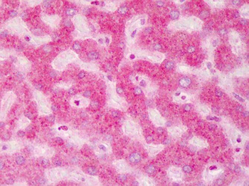
Immunohistochemistry of ABCB5 antibody. Tissue: human liver. Fixation: formalin fixed paraffin embedded. Antigen retrieval: user optimized. Primary antibody: ABCB5 at 1:200. Secondary antibody: Peroxidase goat anti-rabbit at 1:10000 for 45 min at RT. Localization: Moderate to strong cytoplasmic and membranous staining was observed in hepatocytes. Occasional nuclear staining was observed in hepatocytes and sinusoidal cells. Staining: antibody as precipitated red signal with a hematoxylin purple nuclear counterstain.
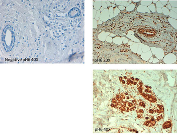
Immunohistochemistry of Rabbit anti ABCB5 antibody. Tissue: human breast carcinoma at pH6. (left, neg control) (right, 20X, 40X). Fixation: formalin fixed paraffin embedded. Primary antibody: ABCB5 antibody at 10 µg/mL for 1 h at RT. Secondary antibody: Peroxidase rabbit secondary antibody at 1:10000 for 45 min at RT. Localization: ABCB5 is cytoplasmic. Staining: ABCB5 as precipitated brown signal with hematoxylin purple nuclear counterstain.
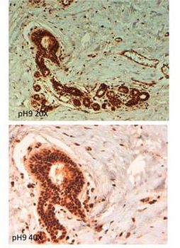
Immunohistochemistry of Rabbit anti ABCB5 antibody. Tissue: human breast carcinoma at pH9. (20X, 40X) Fixation: formalin fixed paraffin embedded. Primary antibody: ABCB5 antibody at 10 µg/mL for 1 h at RT. Secondary antibody: Peroxidase rabbit secondary antibody at 1:10000 for 45 min at RT. Localization: ABCB5 is cytoplasmic. Staining: ABCB5 as precipitated brown signal.
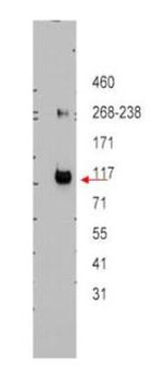
Western blot using Biorbyt's affinity purified anti-ABCB5 antibody shows detection of ABCB5 beta in ~12.5 ug of transfected-Hi5 whole cell lysate. No reaction was seen when antibody was pre-incubated with the immunizing peptide (data not shown). A 3-8% Tris-acetate gel was used for separation. The arrowhead corresponds to 117 kDa ABCB5. The membrane was probed with the primary antibody at a 1:10000 dilution in 5% milk in TBST at 4°C, overnight.
- Item 1 of 7
ABCB5 Antibody [orb570329]
ELISA, FC, ICC, IF, IHC, WB
Human, Mouse, Rat
Rabbit
Polyclonal
Unconjugated
10 μg, 100 μg - Item 1 of 6
- Item 1 of 4
- Item 1 of 3
- Item 1 of 3


Submit a review
Filter by Rating

- 5 stars

- 4 stars
- 3 stars
- 2 stars
- 1 stars
